Newborn Congenital Heart Disease, Part 2

Newborn Congenital Heart Disease, Part 2

In Part 2 of this series on newborn congenital heart disease, we will review the evaluation of a newborn who presents to the emergency department.  Check out Part 1 of this series, which reviewed the presentation of a newborn with suspected congenital heart disease.

Evaluation

In addition to your usual thorough exam of a newborn, below are some additional tools to include in your evaluation.

Systolic Blood Pressure Differential

If the right upper extremity and either lower extremity have a systolic measurement greater than 10 mmHg difference, you should consider CHD, in particular coarctation of the aorta.

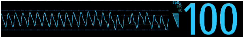
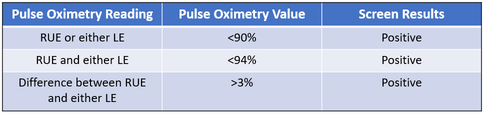
PreDuctal and PostDuctal Saturations

Place a pulse oximeter on the right upper extremity and either lower extremity. The table below will help you interpret the results.

If you have a positive screen, consider CHD.  

Chest X-Ray

Boot Shaped Heart of Tetralogy of Fallot
Snowman Heart of Total Anomalous Pulmonary Venous Return
Egg on a String Heart of Transposition of the Great Vessels

The images above are the classic chest x-ray images of specific congenital heart lesions.   However, remember your chest x-ray may be normal, and this does not rule out CHD.

Images obtained from this free to use source.

EKG

Obtaining an EKG to evaluate for axis deviation can be helpful for evaluation.  Right axis deviation suggests strong rightward forces like single ventricle physiology (HLHS) or right outflow tract obstructive lesions such as tricuspid atresia, pulmonary atresia, or pulmonary stenosis.

Extreme right axis deviation suggests an AV canal defect. 

References

Strobel, A. M., & Lu, L. N. (2015). The critically ill infant with congenital heart disease. Emergency Medicine Clinics33(3), 501-518.

Katie Edmunds is a current Pediatric Emergency Medicine Fellow interested in creating PEM 4 all those who are given the responsibility of treating the sickest babies wherever they may be.

Leave a Reply