Not so FAST– Pitfalls of POCUS FAST

Not so FAST– Pitfalls of POCUS FAST

This PEM 4 Breakdown summarizes the highlights of our most recent ultrasound conference.  With help from our ultrasound guru Dr. Ted Brenkert, we will discuss pitfalls of the FAST exam and how to improve your image acquisition.  Thanks to Dr. Brenkert for providing us these images and guidance on how to make them better!!

Pitfall #1

Here is an image of the RUQ. What is the problem with this image, and what do you need to do to improve your view?

If you need a hint, here is the LUQ view with the same problem!

The probe is too anterior in this RUQ and LUQ view.  

The fix here is to sweep or slide the probe more posterior (towards the bed).  In this LUQ image, you can now see both the spleen and kidney to evaluate the splenorenal recess, spleen tip, and the subphrenic space.  The same fix applies to the RUQ view.


Pitfall #2

An images of the RUQ is below.  Again-  what is the issue with this image, and what is the fix?

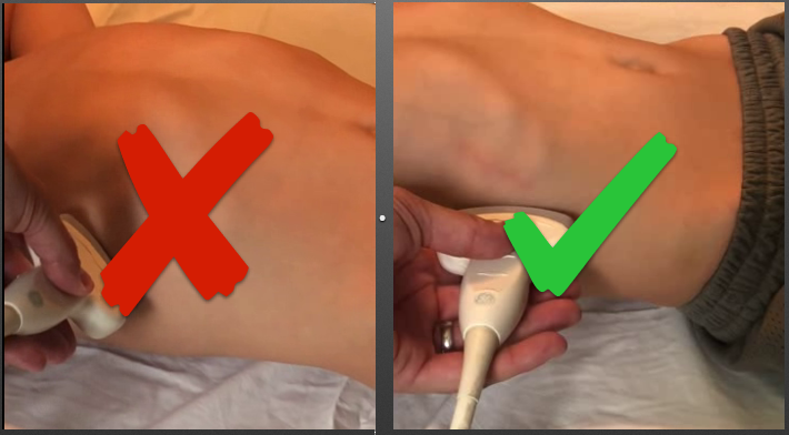
This image is too rotated, which is not allowing us to completely evaluate the hepatorenal junction.

Remember- keep the probe parallel with the bed when obtaining FAST images!

Pitfall #3

How about these RUQ and LUQ images- what are we missing here?

The inferior tip of the liver in the RUQ view and the inferior renal pole in both of the views are missing.

Pitfall #4

Our final FAST Pitfall- what is the problem with this image of the bladder?

There is too much posterior acoustic enhancement, obscuring the imaging behind the bladder.

Use TGC (Time Gain Compensation) to decrease the gain behind the bladder. (Forgive the incorrect labeling of the image!)

Katie Edmunds is a current Pediatric Emergency Medicine Fellow interested in creating PEM 4 all those who are given the responsibility of treating the sickest babies wherever they may be.

Leave a Reply